
Living with oligodendroglioma can be challenging, but you are not alone. In this section, we’ll talk about common symptoms and share tips on how to manage them day-to-day.
Headaches and seizures are common. Most patients also notice changes in their ability to concentrate, make decisions, and stay motivated: the executive functions. Our goal is to give you and your loved ones information that can help you make better choices to make everyday life a little easier.
Headaches

It’s normal to feel concerned if you experience headaches after a brain tumor diagnosis — but in most cases, headaches can be managed with the right care and support.
In another section, we talk about headaches in people who are worried they might have a brain tumor but haven’t been diagnosed.
You can find that information HERE.
This section focuses on headaches in people who have already been diagnosed with, and treated for, a brain tumor — and how they can manage their headaches in practice.
Managing Headaches After a Brain Tumor Diagnosis
Having a brain tumor doesn’t protect you from other types of headaches. In fact, headaches are very common.
About half of all adults have recurring headaches, and 9 out of 10 people have experienced a headache at least once.
Before we can choose the right treatment, it’s important to understand what kind of headache you’re dealing with. Different types of headaches often need different kinds of care.
Headaches are usually divided into the following types:
- Migraine and its subtypes
- Tension-type headaches
- Medication overuse headaches
- Other headaches (for example, those linked to infections like sinusitis, or following injuries)
In the next section, we’ll briefly review the most common types of headaches and share how they can be treated effectively.
More detailed information about headaches can be found for example on the homepages of NIH https://www.ninds.nih.gov/health-information/disorders/headache
Below, we briefly discuss the most common types of headaches and provide information about their treatments.
Is there a specific type of headache related to brain tumors in patients who have already been diagnosed and treated for a brain tumor?
Based on experience, this seems to be quite rare. People who have had a brain tumor often experience migraines and tension-type headaches just like others of the same age group.
In some cases, surgery to remove a brain tumor can affect the muscles used for chewing on one side of the face and temple region. This can lead to problems with how the teeth meet (the “bite”), causing one-sided headaches that can be bothersome and may need treatment.
If the tumor has caused vision problems or hearing loss on one side, a person might need to tilt their head to compensate. Over time, this can lead to tension in the neck muscles and cause headaches.
Sometimes migraines may worsen after a brain tumor diagnosis. On the other hand, steroid treatments (such as those given during radiation therapy) can completely relieve headaches. When the steroid treatment is stopped, headaches might return.
This may cause concern that the tumor is growing again, but most of the time, the headaches simply return because the steroid’s effects have worn off — not because of tumor growth.
Steroids can also be used to break a difficult cycle of headaches, even when there is no tumor involved. Neurologists know from experience that steroids can help relieve severe headaches. However, because steroids can cause side effects, they are usually considered a last-resort treatment option for persistent headaches.
Types of headaches
Migraine
A The patient has had at least 5 attacks of headache that fulfill the criteria B-D.
B A migraine headache attack lasts typically (without medication) between 4 and 24 hours . The headache has the following C features:
- Moderate to severe pain,
- Throbbing or pulsating pain,
- Pain is on one side of the head, and
- Physical activity (such as walking) worsens the pain.
At least two of these C features must be present.
Additionally, D features include:
- Sensitivity to light and/or sound, and/or
- Nausea and vomiting.
Patients may not experience all of these symptoms. However, if at least two C features and one D feature are present, the headache can usually be classified as a migraine.
Often migraines run in families. It’s important to know that some people experience migraine attacks very rarely. For example, someone might have a migraine once a year without warning symptoms (as occurs in about 85% of cases) and may not even realize they have migraines. Therefore, a lack of clear family history doesn’t rule out migraine.
Migraines usually begin before the age of 40. However, some brain tumor patients may experience their first migraines after their diagnosis and treatment.
If you treat a migraine right at the start — for example, by taking two effervescent paracetamol tablets (total 1000 mg), ibuprofen, aspirin, or a combination of these with caffeine — it can often help. Dissolvable tablets in water may be absorbed slightly faster than regular tablets.
These medications are less effective if taken after the headache is already pounding strongly.
Today, new targeted medications are available for migraines, such as different types of triptans. If headaches are difficult to manage, it’s a good idea to talk with your doctor or ask for a referral to a neurologist.

Tension-Type Headache
Tension-type headaches are steady, non-pulsating headaches that often get worse during the afternoon. Physical activity usually eases the pain.
Unlike migraines, tension headaches typically do not cause nausea or sensitivity to light or sound.
They tend to respond less well to medication compared to migraines.
Tension headaches may also cause sharp, stabbing pains felt on the scalp, often around the surgical area, especially in patients who have had radiation therapy. In these cases, muscles and scar tissue may be irritated and sensitive to touch.
A short course of anti-inflammatory medication might help. Physiotherapy can also be useful — but it’s important to wait until the tissues, including skin and muscles, have fully healed after cancer treatments before starting physiotherapy.
Sometimes but very rarely, the wound may get infected. Usually the scar gets read, sensitive to touch, maybe also warm and may exude pus. This is rare, but possible. Then the pain is actually not headache but sucll and scalp ache. It needs immediate medical attention. Usually the skin has to be opened so that all pus can be drained and cleansed and then an antibiotic started at the health providers discernment.
Problems with the bite (how the teeth meet) can also cause headaches. If chewing muscles are damaged during surgery (usually on one side), it may lead to bite problems and headaches. These can be very bothersome. A dentist -or physiotherapist- specialized in bite issues can offer help if needed.
Medication-Overuse Headache
Brain tumor patients are not immune to medication-overuse headaches. If headaches are severe, it’s easy to fall into the habit of taking painkillers frequently.
It is estimated that if medication is used on more than one-third of days, there’s a risk of developing medication-overuse headaches — in addition to the original headache problem.
The only real treatment for medication-overuse headaches is stopping the painkillers. Sometimes this can be challenging and requires support from a doctor experienced in headache management. Fortunately, this is a rare problem among brain tumor patients.
New, Sudden-Onset Headaches
If a brain tumor patient develops a new, sudden headache, it’s important to investigate the cause.
Doctors will want to rule out bleeding in the tumor area — which is possible but very rare — or an infection such as meningitis (also very rare).
These conditions are typically associated with neck stiffness, new neurological symptoms, and in the case of infection, fever.
Bleeding headaches start within minutes; infection-related headaches develop over a few hours.
If you experience a sudden, severe new headache, you should seek medical attention immediately.
Päänsärky etenevässä kasvaimessa
Headache with a Growing Tumor
If a brain tumor has returned and continues to grow despite treatment, about one-third of patients may experience headaches.
This type of headache is often treated with corticosteroids because the pain is usually related to swelling caused by the tumor’s progression.
Anti-inflammatory pain medications can also be used, as well as medications typically used for nerve pain, such as certain antidepressants that raise the pain threshold, and sometimes stronger painkillers if needed.
Each situation needs to be assessed individually, and usually a brain scan is needed to check what’s happening.
What does the doctor ask during a headache consultation?
What Will the Doctor Ask You at a Headache Appointment?
If you are going to see a doctor about your headaches, it’s helpful to think ahead about answers to the following questions:
- When did the headache start? Hours, days, weeks, months, or even years ago? Has it changed over time?
- How severe is the pain? Try rating it on a scale from 0 to 10, where 0 means no pain at all and 10 means the worst pain imaginable.
For example:- 8/10 is very painful but could still get worse.
- 3–4/10 is noticeable but stays more in the background.
It’s often hard to give a precise number, but this scale gives the doctor a clearer idea of the severity.
- Does the pain interfere with your daily life?
- Does it force you to stop all activities and rest?
- Or can you push through it with some effort?
- How would you describe the pain?
Burning, stabbing, throbbing, sharp, tearing, heavy, pounding, pressing, or wave-like? Or something else? - Is the pain constant or does it vary?
- What makes it worse?
- What makes it better?
- Where exactly in your head do you feel it?
- Which medications have helped? Which haven’t?
- Are there any other symptoms along with the headache? Such as visual disturbances, nausea, etc.
- Are there different types of headaches or just one type with varying intensity?
Thinking about these questions ahead of time — and even writing down the answers — can help your doctor understand your situation more quickly and plan your treatment more efficiently.

Practical Tips
From experience, it’s harder for doctors to help when they get vague answers.
For example, if the doctor asks, “Is the pain severe and how often does it happen?” and the answer is simply “It’s bad, and it happens a lot,” it’s not very informative.
It’s much clearer if you say, “It’s quite strong, happens about twice a week, started around late autumn, and I always feel nauseous when it hits.”
That gives the doctor a good foundation to build on with further questions.
.
One useful tip:
If possible, let the doctor ask their questions first before you offer your own idea of the diagnosis.
If you immediately say, “This feels just like my usual migraine,” before the doctor has asked anything, it might complicate things.
The doctor needs to get a full picture before making any judgment, so let him/her gather the information first — then discuss together what the cause might be.
Another helpful thing to share with your doctor is what is your main concern:
- Is it the pain itself that worries you most?
- Or are you more worried about what is causing the pain?
Some patients feel they can tolerate the pain but are anxious about whether it signals tumor growth.
Others may think, “It doesn’t matter what’s causing it — I just want relief.”
If you know what matters most to you, tell your doctor.
It will help you both understand each other better and find the best approach.
I have seen situations in which the patient is primarily troubled by the pain, while the doctor focuses their skills and efforts on finding its cause. Conversely, a doctor may work hard to relieve the symptoms without realizing that the patient wants to know why the pain is occurring. In such cases, it can be difficult to reach a mutual understanding unless the situation is explicitly put into words.
Epilepsy
Up to 70% of patients with brain tumors experience an epileptic seizure during the course of their illness. Seizures are the most common symptom leading to the diagnosis of a brain tumor in adults. An epileptic seizure is a temporary electrical disturbance in the brain. It is a symptom of the tumor, not a separate disease. The following sections explain the nature of seizures and their treatment in more detail.
What is epilepsy?
Epilepsy is a predisposition to recurrent seizures. According to current classification criteria, a single seizure is sufficient for a diagnosis of epilepsy if the risk of another seizure due to a brain disorder is greater than 60%. In patients with brain tumors, one seizure is generally sufficient to establish a diagnosis of epilepsy.
Seizures are more common in patients whose tumors are slow-growing, or low-grade, and located in the cerebral cortex.
In patients with brain tumors, seizures may be associated with abnormal cells that have developed in the tissue surrounding the tumor. They may also result from an imbalance of chemical substances in the brain caused by the tumor. Both mechanisms can disrupt the brain’s normal electrical activity.
Most patients with brain tumors experience at least one seizure. However, this may be the only seizure they ever have, or seizures may occur only for a short period, for example, due to brain swelling after surgery. High doses of corticosteroids may increase the likelihood of seizures. Nevertheless, only about 10% of brain tumor patients develop seizures later in the course of their illness if they did not experience them at the time of diagnosis. In patients with low-grade gliomas, such as oligodendrogliomas, the recurrence of epileptic seizures does not usually indicate that the tumor has returned.
What is a seizure?
Our brains contain billions of nerve cells that regulate the functions of the body and are also responsible for thinking, emotions, sensory functions, and their regulation, among other things. Nerve cells communicate with one another through electrical signals. Electrical activity is constantly occurring in the brain.
When abnormal electrical activity suddenly occurs in an area of the brain, it may involve excessive or synchronized electrical discharges across an area of varying size. This produces a seizure, disrupting the brain’s normal functioning and causing various symptoms. It is similar to a short circuit in an electrical system.
Types of seizures
When people talk about an epileptic seizure, they most often think of a convulsive seizure in which a person loses consciousness, the body becomes stiff, the person falls to the ground, and the limbs jerk involuntarily.
Brain tumors can cause many different types of seizures. Symptoms may range from a brief episode lasting a few seconds to a few minutes, during which a person experiences an unusual sensation or appears absent, to seizures involving loss of consciousness and convulsions. The latter are known in medical terminology as tonic-clonic seizures. Tonic means stiffening, while clonic refers to involuntary jerking movements.
No single pattern of seizure symptoms is characteristic of brain tumors. The symptoms that occur during a seizure depend on the location of the abnormal electrical discharge in the brain and how far it spreads.
Epilepsia – Epilepsy
Jopa 70%: lla aivokasvainpotilaista on epileptinen kohtaus tautinsa aikana. Kohtaukset ovat yleisin aivokasvaimen toteamiseen johtava oire aikuisilla. Epileptinen kohtaus on ohi menevä sähköhäiriö aivoissa. Se on kasvaimen oire, ei erillinen tauti. Seuraavassa selitetään kohtausten laatua ja hoitoa tarkemmin.
Mikä on epilepsia? – What is epilepsy?
Epilepsia on taipumus toistuviin kohtauksiin. Nykyluokituksen mukaan yksi kohtaus riittää epilepsia-diagnoosiin, jos todennäköisyys saada toinen kohtaus aivotaudin vuoksi on yli 60%. Yleensä aivokasvainpotilaalla riittää yksi kohtaus epilepsiadiagnoosiin.
Kohtauksia on enemmän niillä, joiden kasvain on hitaasti kasvava eli matala-asteinen ja kasvain sijaitsee aivokuorella.
Aivokasvainpotilailla kohtaukset voivat liittyä kasvaimen ympärillä oleviin soluihin, jotka ovat kehittyneet epänormaalisti. Ne voivat myös johtua kasvaimen aiheuttamasta kemiallisten aineiden epätasapainosta aivoissa. Nämä molemmat voivat häiritä aivojen normaalia sähköistä toimintaa.
Suurimmalla osalla aivokasvainpotilaista on ainakin yksi kohtaus. Tämä voi kuitenkin jäädä ainoaksi tai niitä voi olla vain lyhyen ajan, esimerkiksi aivojen turvotuksesta johtuen leikkauksen jälkeen. Suuret kortisoniannokset voivat lisätä kohtaustodennäköisyyttä. Kuitenkin vain noin 10%lle aivokasvainpotilaista tulee kohtauksia myöhemmin taudinkulussa, ellei niitä ole ollut jo toteamisvaiheessa. Matala-asteista glioomaa sairastavilla, kuten oligodendroglioomapotilailla epileptisten kohtausten alkaminen uudelleen ei yleensä merkitse kasvaimen uusiutumista.
Mikä on kohtaus? – What is a seizure?
Aivoissamme on miljardeja hermosoluja, joiden avulla elimistön toimintaa säädellään ja joiden toiminnasta muodostuu myös ajattelu, tunteet, aistitoiminto ja niiden säätely jne. Hermosolut viestivät keskenään sähkösignaaleilla. Aivoissamme on sähköistä toimintaa jatkuvasti.
Kun yhtäkkiä on epänormaaleja, poikkeavia joko liiallisia tai samanaikaisia sähköisiä toimintoja laajuudeltaan vaihtelevalla aivoalueella, tulee kohtaus. Tämä sekoittaa aivojen normaalia toimintaa. Sekaannus aiheuttaa erilaisia oireita. Tämä on kuin oikosulku sähkötoiminnassa.
Kohtausten lajit
Kun puhutaan epileptisestä kohtauksesta, useimmiten ajattellaan kouristuskohtausta, jossa ihminen menettää tajuntansa, keho jäykistyy, henkilö vaipuu maahan ja raajat nykivät.
Aivokasvain aiheuttaa monenlaisia kohtauksia. Ne voivat vaihdella hetkittäisestä, muutaman sekunnin – muutaman minuutin omituisesta tai poissaolevasta olosta tajuttomuus-kouristuskohtauksiin, joita kutsutaan lääkärilatinaksi toonis-kloonisiksi. Tooninen tarkoittaa jäykkää ja klooninen tahattomasti nykivää. Mikään yksittäinen kohtausoireisto ei ole aivokasvaimelle tyypillinen. Poikkeavan sähköpurkauksen sijainnista ja leviämisalueesta aivoissa riippuu, minkälaisia oireita kohtauksen aikana ilmaantuu.
Treatment of Epilepsy
In patients with brain tumors, epilepsy is also affected by the various treatments used for the tumor. Surgery may reduce epileptic seizures, as may radiotherapy. Corticosteroids can reduce seizures by reducing swelling. However, it is known that high doses of corticosteroids can also trigger seizures.
Otherwise, epilepsy associated with brain tumors is treated in the same way as other forms of epilepsy, as there are few specific recommendations for this patient group. Last year, a group of neuro-oncologists in Italy developed a framework of recommendations based on a comprehensive literature review. I will refer to these recommendations here where relevant. A reference is provided at the end of the text for those interested in reading the article (Maschio M et al.).
Antiepileptic Medications
Epilepsy is treated with medications intended to prevent seizures from recurring. Because seizures are common in patients with brain tumors, some have argued that medication should be used to prevent seizures even in patients who have never experienced one. However, the available studies and investigations have led to the well-founded conclusion that preventive, or prophylactic, medication is not beneficial. In other words, if a patient has not experienced seizures, medication is not prescribed. The Italian group also reached this recommendation (Maschio M et al.).
Medication is usually started after the first seizure, unless there is a specific reason to withhold treatment.
There are many antiepileptic medications that work in different ways, and treatment is selected individually. Many medications are introduced by gradually increasing the dose to the target level in order to minimize adverse effects. In patients with tumors, however, the dose often needs to be increased more rapidly because another seizure may occur.
If one medication is insufficient to prevent seizures, two or more medications with different mechanisms of action can be used together. Approximately 30% of patients need more than one medication. In general, more than three different antiepileptic medications should not be used simultaneously. In such situations, other treatment options should be considered.
Medication dosing requires careful management and is partly adjusted based on blood concentrations measured in laboratory tests. However, the current principle is primarily to listen to the patient and assess how they are feeling, rather than treating laboratory results alone.
There are some special considerations when selecting medications for patients with brain tumors. However, there is no research evidence demonstrating that particular medications are more effective for specific tumor types or particular seizure types in patients with brain tumors. In general, a medication should be effective, have few side effects that are tolerable for brain tumor patients, and not interact with other medications used to treat the tumor, such as chemotherapy drugs. If possible, it should also help inhibit tumor growth.
Some antiepileptic medications are eliminated from the body through liver metabolism. Certain medications enhance, or induce, the activity of liver enzymes involved in this process. A problem can arise if the increased enzyme activity also causes medications intended to treat the tumor to be broken down and eliminated from the body too quickly.
For this reason, medications that do not induce liver enzymes are recommended. The most common enzyme-inducing medications are carbamazepine, phenytoin, and phenobarbital. Phenytoin and phenobarbital are rarely used in Finland. If no medications are being used to treat the tumor, enzyme induction is naturally not a concern.
Small studies have suggested that patients receiving valproate or levetiracetam may have a better prognosis. However, this has not been conclusively confirmed. It is therefore not possible to state with confidence that any antiepileptic medication improves the prognosis of the tumor itself.
When it comes to adverse effects, it is worth remembering that some people experience them while others do not. We know too little to predict who will develop these effects. Reading the list of adverse effects for any medication can be alarming, because even the rarest effects are included. In practice, most individual side effects occur in only a small proportion of patients, often just a few out of every hundred. One doctor advised that it is best not to read these lists of side effects UNLESS something unusual occurs after starting the medication.
When a doctor prescribes medication, they are aware of its potential adverse effects. All medications have side effects, some more than others. These effects do not come as a surprise to the doctor; they have already been considered. When receiving a new medication, it may be helpful to ask the doctor which side effects are common. The doctor will usually explain this to the patient anyway.
Epilepsian hoito – Treatment of epilepsy
Aivokasvainpotilailla epilepsiaan vaikuttavat myös kasvaimen eri hoidot. Leikkaus voi vähentää epileptisiä kohtauksia, samoin sädehoito. Kortisoni voi vähentää kohtauksia, kun se vähentää turvotusta. Suurista kortisoniannoksista tiedetään kuitenkin, että ne voivat laukaista kohtauksia.
Muuten aivokasvaimiin liittyvää epilepsiaa hoidetaan kuten muitakin epilepsioita, koska erityisiä suosituksia ei juuri ole. Italiassa neuro-onkologien ryhmä on tehnyt viime vuonna perusteellisen kirjallisuuskatsauksen perusteella suositusrunkoa. Viittaan siihen tässä siinä määrin, kuin se vaikuttaa tarpeelliselta. Tekstin lopussa on viite, jonka perusteella artikkeli löytyy niille, jotka ovat suosituksesta kiinnostuneet. (Maschio M ym. )
Epilepsialääkkeet
Epilepsiaa hoidetaan lääkkeillä, joiden tarkoitus on estää kouristuskohtausten uusiutuminen. Koska kohtaukset ovat kasvainpotilailla yleisiä, ovat toiset ajatelleet, että kohtauksia kannattaa estää lääkkeillä, vaikka ei mitään kohtauksia olisi ollutkaan. Kaikkien tehtyjen tutkimusten ja selvitysten perusteella on kuitenkin muodostunut perusteltu käsitys, että ennalta ehkäisevä eli profylaktinen lääkitys ei auta. Jos ei ole ollut kohtauksia, ei siis anneta lääkitystäkään. Tähän suositukseen päätyy myös italialaisten ryhmä (Maschio M ym. )
Lääkitys aloitetaan yleensä ensimmäisen kohtauksen jälkeen ellei ole jotakin erityistä syytä pidättäytyä lääkityksestä.
Epilepsialääkkeitä on monin eri tavoin vaikuttavia, ja lääkitys valitaan yksilöllisesti. Useat lääkkeet aloitetaan nostamalla annosta hitaasti tavoitetasolle haittavaikutusten välttämiseksi. Kasvaimen kyseessä ollen, joudutaan usein nostamaan annosta ripeästi, koska on mahdollista, että kohtaus uusii.
Jos yksi lääke ei riitä pitämään kohtauksia poissa, voidaan käyttää yhtä aikaa lääkkeitä, joilla on erilainen vaikutusmekanismi. Noin 30 % potilaista joutuu käyttämään useampaa kuin yhtä lääkettä. Normaalisti yli kolmea eri epilepsialääkettä ei pitäisi käyttää yhtä aikaa, vaan tällaisissa tilanteissa on harkittava muita hoitovaihtoehtoja. Lääkkeiden annostelu on tarkkaa ja sitä säädellään osin laboratoriossa tutkittujen lääkkeiden veripitoisuuksien perusteella. Nykyään periaatteena on kuitenkin pääasiassa kuunnella potilasta ja hänen vointiaan eikä hoitaa pelkkiä laboratoriokoetuloksia.
Aivokasvainpotilaiden lääkevalinnassa on joitakin erityispiirteitä. Ei kuitenkaan ole mitään tutkimusnäyttöä siitä, että joku tai jotkut lääkkeet olisivat tiettyihin kasvaimiin parempia tai tietyntyyppisiin kohtauksiin aivokasvainpotilailla parempia. Yleisesti lääkkeen pitäisi olla tehokas, sivuvaikutukset vähäisiä ja aivokasvainpotilaille siedettäviä, lääke ei saisi aiheuttaa yhteisvaikutuksia muiden kasvaimen hoitoon käytettävien lääkkeiden, kuten sytostaattien kanssa ja jos mahdollista, sen pitäisi osaltaan hillitä kasvaimen kasvua.
Osa epilepsialääkkeistä poistuu elimistöstä maksan aineenvaihdunnan kautta. Osa lääkkeistä tehostaa eli stimuloi maksan entsyymien toimintaa tässä prosessissa. Ongelmaksi voi muodostua, jos tehostunut maksan toiminta käsittelee myös kasvaimen hoitoon tarkoitetut lääkkeet nopeasti pois elimistöstä. Siksi suositellaan sellaisia lääkkeitä, jotka eivät stimuloi maksaentsyymejä. Yleisimmät tällaiset entsyymejä stimuloivat lääkkeet ovat karbamatsepiini, fenytoiini ja fenoarbitaali. Fenytoiinia ja fenobarbitaali ei Suomessa juurikaan käytetä. Mikäli kasvaimen hoitoon ei käytetä lääkkeitä, ei entsyymistimulaatiolla luonnollisesti ole merkitystä.
Pienissä tutkimuksissa on esitetty, että valproaattia tai levetirasetaamia saavilla potilailla olisi parempi ennuste. Tätä ei kuitenkaan ole kyetty varmistamaan. Ei voida siis vakuuttavasti todeta, että mikään epilepsialääke parantaisi kasvaimen ennustetta.
Haittavaikutuksista kannattaa muistaa, että joku saa sivuvaikutuksia, toinen ei. Tiedämme tästä liian vähän, jotta voisimme päätellä, kuka näitä sivuvaikutuksia saa. Jos lukee minkä tahansa lääkkeen sivuvaikutusluettelon, löytää miltei mitä tahansa, koska kaikkein harvinaisimmatkin sivuvaikutukset on mainittu. Yleensä niitä tulee vain muutamalle potilaalle sadasta. Eräs lääkäri suositteli, ettei näitä sivuvaikutusluetteloita kannata lukea PAITSI jos lääkkeen aloituksen jälkeen ilmenee jotakin outoa.
Kun lääkäri määrää lääkettä potilaalle, hän on tietoinen sivuvaikutuksista. Kaikilla lääkkeillä on sivuvaikutuksia, joillakuilla enemmän, toisilla vähemmän. Sivuvaikutukset eivät tule lääkärille yllätyksenä, niitä on pohdittu. Kannattaa ehkä kysyä lääkäriltä, kun saa uutta lääkettä, mitkä ovat yleisiä sivuvaikutuksia. Lääkäri yleensä jo kertookin tästä potilaalle.
Suomalainen lääkäriseura Duodecim ylläpitää Potilaiden terveyskirjastoa ja siellä on Lääketietokeskuksen potilaiden lääkeopas miltei parista tuhannesta lääkkeestä. Sen löytää tekemällä haun: Lääkeopas Terveyskirjasto.
The Most Common Medications for Brain Tumor-Related Epilepsy
The Italian experts recommend levetiracetam, lamotrigine, topiramate, and valproate. In Finland, oxcarbazepine is also frequently used, although it is a mild enzyme inducer. Zonisamide, lacosamide, perampanel, and clobazam may also be considered, particularly when seizures continue despite medication and a second or third drug is added.
The most commonly used antiepileptic medication in patients with brain tumors today is probably levetiracetam (Keppra®, Kevesy®, Levetiracetam®, Matever®). It has no drug interactions. Side effects are uncommon, and it is generally well tolerated. However, particular attention should be paid to patients with frontal lobe tumors, as levetiracetam can cause various symptoms, mainly affecting emotional regulation, such as irritability or anxiety.
In my own experience, there was a middle-aged mother whose levetiracetam had to be changed because she became so irritable that her family could no longer cope with her behavior. After the medication was changed, the situation settled down. There are many similar stories.
Another advantage of levetiracetam is that its blood levels do not need to be monitored through laboratory tests, nor are laboratory tests otherwise necessary. However, in some situations, it is not sufficiently effective.
A very recent derivative of levetiracetam is brivaracetam (Briviact®). It is as effective as levetiracetam but has fewer side effects. Emotional regulation problems have also been reported less frequently than with levetiracetam. In Finland, brivaracetam is eligible for both basic and special reimbursement. Special reimbursement applies to this medication if the patient already qualifies for it with levetiracetam.
Oxcarbazepine (Apydan®, Oxcarbazepin®, Trileptal®) is well tolerated and easy to take. However, it has two relatively common and sometimes troublesome side effects.
It can lower the sodium concentration in the blood. The body can generally adapt to this because the decrease occurs gradually. Blood sodium levels do not usually need to be monitored, as a small, gradual decrease below the normal range is not dangerous. However, sodium levels should not fall below 125 mmol/L.
This can sometimes cause problems, particularly during hot summer weather, when salt is lost through sweating. Ordinary table salt, or sodium chloride, can be added to food to increase sodium levels, but this is not a sustainable solution to the problem. Sometimes the medication needs to be changed for this reason. Naturally, the situation must be assessed clinically on an individual basis.
Another sometimes troublesome side effect of oxcarbazepine is brain fatigue. This is less well recognized. Because the tumor itself can also cause fatigue, medication-related fatigue may not be identified. However, fatigue can be significantly disabling, so this is not a minor problem for the patient. Many other medications are now available, so it is worth discussing this problem with the treating physician and asking whether the medication could be changed.
For example, lamotrigine and valproate rarely cause fatigue. Topiramate, on the other hand, can cause fatigue and slow cognitive function. In Finland, it is used less frequently for epilepsy associated with brain tumors.
Valproate (Absenor®, Deprakine®, Orfiril®, Valproat®) is used fairly often to treat brain tumor-related epilepsy. It may slow the growth of some tumors. However, this has not been studied sufficiently to establish this with certainty. It is also not known which types of tumors might be affected or what dose of valproate might be required to inhibit their growth.
Valproate cannot be used during pregnancy because it causes birth defects. Pregnancy is relatively uncommon among patients with brain tumors.
A significant problem is weight gain. The mechanisms by which valproate affects weight are also poorly understood. Based on my experience, the effect is not simply due to increased appetite; valproate also affects metabolism.
Lamotrigine (Lamictal®, Lamotrigin®) is a good medication for many people. One possible adverse effect is a skin reaction, which is unusual in that it is dose-dependent.
This differs from allergic reactions, in which a rash may eventually occur even with a very small dose. With lamotrigine, the situation is different. As one patient described it:
“Either I take more medication and remain seizure-free (my seizures are mainly brief, focal episodes involving strange sensations), but suffer from a rash, or I reduce the dose, in which case the rash disappears but the seizures return.”
First Aid for a Convulsive Seizure
In patients with brain tumors who are already taking antiepileptic medication, seizures are usually brief and self-limiting. The first thing to do is check the time so that you know how long the seizure lasts.
If the seizure lasts less than five minutes, there is no need to call an ambulance, according to the guidance in this text. If the situation resolves spontaneously and the seizure does not recur, it may be sufficient to contact the patient’s treating unit during office hours.
If anything is unclear or you have questions, you can call the emergency department of the treating hospital or the nearest central hospital, preferably its neurology department. At the beginning of the call, it is worth explaining what type of tumor the patient has, which medication they are taking, and whether there was any particular factor that may have triggered the seizure. Such factors may include forgetting to take medication, sleep deprivation, or a gastrointestinal illness.
During a convulsive seizure, proceed as follows:
Protect the person from injury: Make sure they do not hit themselves against nearby objects, and keep the surrounding area calm.
Do not put anything in their mouth, for example, to prevent them from biting their tongue.
Place the person in the recovery position once the convulsions have stopped, to prevent them from inhaling saliva or other fluids.
Call the emergency number (112) if the convulsions last longer than five minutes or if another seizure occurs.
After a convulsive seizure in a person with epilepsy has ended, make sure the person is fully conscious before leaving them.
Yleisimmät lääkkeet aivokasvaimen aiheuttamaan epilepsiaan
Italialaiset päätyvät suosittelemaan levetirasetaamia, lamotrigiiniä, topiramaattia ja valproaattia. Suomessa käytetään usein myös okskarbatsepiiniä, joka on lievä entsyymistimulaattori. Zonisamidi, lakosamidi ja perampaneeli sekä klobatsaami voivat myös tulla kyseeseen. Erityisesti, jos kohtauksia tulee lääkityksestä huolimatta ja päädytään lisäämään toinen tai kolmas lääke.
Nykyisin yleisin epilepsialääke aivokasvainpotilailla lienee levetirasetaami (Keppra®, Kevesy®, Levetiracetam®, Matever®). Sillä ei ole yhteisvaikutuksia muiden lääkkeiden kanssa. Sivuvaikutukset ovat harvinaisia ja se on hyvin siedetty. Erityisesti otsalohkokasvainpotilailla on kuitenkin huomattava, että levetirasetaami voi aiheuttaa erilaisia, lähinnä tunnesäätelyä häiritseviä oireita, kuten ärtyisyyttä tai ahdistuneisuutta. Omassa kokemuksessani on keski-ikäinen perheenäiti, jonka levetirasetaami piti vaihtaa, kun hän muuttui niin kiukkuiseksi, että perhe ei enää kestänyt häntä. Kun lääke vaihdettiin, tilanne rauhoittui. Samantapaisia tarinoita löytyy monia. Levetirasetaamissa on myös se hyvä puoli, että sen pitoisuuksia ei tarvitse seurata laboratoriokokein eikä muutenkaan tarvitse ottaa laboratoriokokeita. Joissain tilanteissa se ei kuitenkaan ole riittävän tehokas. Aivan uusi levetirasetaamijohdos on brivarasetaami (Briviact®). Se on yhtä tehokas kuin levetirasetaami, mutta sillä on vähemmän sivuvaikutuksia. Myös tunnesäätelyn häiriöitä on kuvattu vähemmän kuin levetirasetaamilla. Lääkkeellä on peruskorvattavuuden lisäksi erityiskorvattavuus. Erityiskorvattavuuden on voimassa tähän lääkeeseen, jos se on jo levetirasetaamilla.
Okskarbatsepiini (Apydan®, Oxcarbazepin®, Trileptal®) on hyvin siedetty ja helppo ottaa. Sillä on kuitenkin kaksi melko yleistä ja joskus hankalaa sivuvaikutusta. Se voi laskea veren natrium-pitoisuutta. Elimistö tottuu tähän kyllä, koska lasku tapahtuu hitaasti. Verikokein natrium-pitoisuutta ei yleensä tarvitse seurata, koska hitaasti tapahtunut vähäinen lasku normaalirajojen alle ei ole vaarallista. Natrium ei kuitenkaan saisi laskea alle 125mmol/l. Joskus tämä aiheuttaa vaikeuksia erityisesti kesähelteellä, jolloin suolaa erittyy hikoillessa. Tavallista keittosuolaa eli natriumkloridia voidaan lisätä ruokaan ja tällöin natriumpitoisuus nousee, mutta tämä ei ole ongelmalle kestävä ratkaisu. Joskus lääke pitää vaihtaa tästä syystä. Tilanne on luonnollisesti arvioitava kliinisesti potilaskohtaisesti.
Toinen, joskus hankala sivuvaikutus okskarbatsepiinistä, on aivoväsymys. Tämä on huonommin tunnettu. Koska kasvaimeen voi muutenkin liittyä väsymystä, ei lääkkeen aiheuttamaa väsymystä välttämättä tunnisteta. Väsymys voi kuitenkin invalidisoida merkittävästi, joten potilaan kannalta ongelma ei ole vähäinen. Nykyisin on olemassa monia muitakin lääkkeitä, joten tästä ongelmasta kannattaa puhua hoitavalle lääkärille. Tällöin voi kysyä lääkevaihdosta.
Esimerkiksi lamotrigiini tai valproaatti väsyttävät harvoin. Sen sijaan topiramaatti voi väsyttää ja hidastaa aivotoimintaa. Suomessa sitä käytetään harvemmin aivokasvaimiin liittyvään epilepsiaan.
Valproaattia (Absenor®, Deprakine®, Orfiril®, Valproat®) käytetään melko usein aivokasvainepilepsian hoitoon. Se saattaa hidastaa joidenkin kasvainten kasvua. Tätä ei kuitenkaan ole riittävästi tutkittu, jotta tämä tiedettäisiin varmasti. Luonnollisestikaan ei myöskään tiedetä minkälaisten kasvainten kasvua ja millä annoksella valproaatti mahdollisesti hillitsisi. Valproaattia ei voi käyttää raskausaikana, koska se aiheuttaa epämuodostumia. Raskaus tulee aivokasvainpotilailla harvoin kyseeseen. Merkittävä ongelma on painon nousu. Miten valproaatti vaikuttaa painonnousuun on myös huonosti tunnettu. Koekemukseni perusteella kyse ei ole pelkästään kasvaneesta ruokahalusta vaan valproaatti vaikuttaa myös aineenvaihduntaan
Lamotrigiini (Lamictal®, Lamotrigin®) on monille hyvä lääke. Haittavaikutuksina lamotrigiinilla voi olla iho-oireita, jotka ovat sikäli erikoisia, että ne ovat annosriippuvaisia. Toisin kuin allergiset reaktiot, joissa ihottumaa tulee lopulta miten pienestä annoksesta tahansa, on tilanne lamotrigiinin suhteen eri. Niin kuin eräs potilas kuvasi tilannetta: joko otan enemmän lääkettä ja olen ilman kohtauksia (lyhyitä, paikallisia, outoja oloja lähinnä) mutta kärsin ihottumasta tai lasken annosta, jolloin ihottuma häviää, mutta kohtauksia tulee.
Kouristuskohtauksen ensiapu – First aid for a seizure
Aivokasvainpotilaalla, jolla on jo epilepsialääkitys, ovat kohtaukset yleensä lyhyitä ja itsestään rajoittuvia. Kelloa on syytä vilkaista ensimmäiseksi, jotta voi tietää, kauanko kohtaus kesti. Jos kohtaus kestää alle 5 minuuttia, ambulanssia ei tarvitse kutsua paikalle. Jos tilanne rauhoittuu itsestään eikä uusi, riittänee, että soittaa hoitavaan yksikköön virka-aikana.
Mikäli tilanteessa on epäselvää tai kysyttävää, voi soittaa hoitavan sairaalan tai lähimmän keskussairaalan (neurologiseen) päivystykseen. Puhelun alussa kannattaa kertoa, minkälainen kasvain potilaalla on ja mitä lääkettä hän käyttää sekä onko joku erityinen tekijä, joka on laukaissut kohtauksen. Tällaisia voivat olla esimerkiksi lääkkeen unohtaminen, valvominen tai vatsatauti.
Kouristuskohtauksen aikana toimitaan seuraavasti:
- Huolehditaan, että henkilö ei kolhi itseään esineisiin ja ympäristö rauhoitetaan
- Suuhun ei saa laittaa mitään (esimerkiksi estämään kieleen puremista).
- Henkilö käännetään kylkiasentoon, kun kouristelu on päättynyt, jottei hän vedä henkeen sylkeä tms.
- Jos kouristelu kestää yli 5 minuuttia tai se uusiutuu, soitetaan hätänumeroon (112).
- Kun epilepsiapotilaan kouristuskohtaus menee ohitse, varmistetaan ennen paikalta poistumista, että henkilö on täysin tajuissaan.
Luuloja ja tietoa epilepsiasta – Myths and facts about epilepsy
| Luulo (Myytti) | Tieto (Fakta) | |
| Yleisyys | Harvinainen aivokasvaimissa | Yleisin oire aivokasvainpotilailla |
| Yleisyys | Harvinainen väestössä | Suomessa yli 50 000 sairastaa epilepsiaa, eli n. 1% väestöstä; kasvaimet ovat epilepsian aiheuttajana harvinaisia |
| Yleisyys | Jos aikuinen saa epilepsian, aina on kasvain taustalla | N. 20%lla aikuisena epilepsian saaneista on taustalla kasvain |
| Laatu | Tauti | Oire |
| Kohtaus | Samanlainen kaikilla | Kohtauksen oireet riippuvat sähköhäiriön laajuudesta ja sijainnista |
| Kohtaus | Kaikki menevät tajuttomiksi ja raajat nykivät | Vain n. 1/3 kohtaus yleistyy tajuttomuuteen ja raajanykinöihin; kohtauksia on monenlaisia |
| Kohtauksen ensiapu | Pitää yrittää pitää potilas paikallaan ja estää puremasta kieleen laittamalla jotain suuhun | Ainoastaan estä potilasta lyömästä itseään ja käännä kyljelleen; älä laita mitään suuhun |
| Hoito | Epilepsiaa ei voi saada kuriin | Usein epilepsia voidaan hoitaa ja aivokasvainpotilailla se useimmiten saadaan ainakin rajoittumaan |
| Hoito | Sädehoito pahentaa kasvaimeen liittyvää epilepsiaa | Sädehoito usein auttaa kasvaimeen liittyvään epilepsiaan |
Italialainen aivokasvaimiin liittyvän epilepsian tutkimusryhmän suositus:
Management of Epilepsy in Brain Tumors Marta Maschio, Umberto Aguglia, Giuliano Avanzini ym. Neurol Sci .2019 Oct;40(10):2217-2234. doi: 10.1007/s10072-019-04025-9.
.
Disturbances in mental performance (higher brain functions)
Classification of mental performance
Understanding Mental Performance
Disturbances in mental performance — also called higher brain function — occur at some level in most brain tumor patients.
Before we go into the symptoms caused by brain tumor-related damage, let’s first review a few basic concepts to help you understand what we are talking about.
What are Higher Brain Functions?
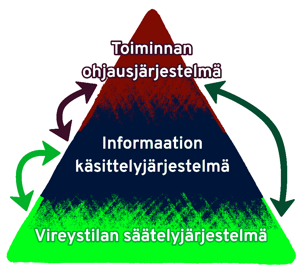
Higher brain functions, or mental performance, can be divided into three functional parts. These functional parts care not localized in a single lobe of the brain but they are dispersed over all parts of the brain.
These divisions aren’t strict, but they give a useful framework:
- Executive Functioning System
- Information Processing System
- Vigilance upholding System
At the core of these functions are highly complex networks of nerve cells, connected by hubs all over the brain.
(1) EXECUTIVE FUNCTIONS, also known as the executive system, operates by programming, regulating, and verifying mental activity. This part of the brain’s neural networks directs, adjusts, and monitors how other neural networks and the entire body are utilized. In practice it helps us plan, control, and check our thoughts and actions. This system plays a key role for example in how we focus, make decisions, solve problems, and control impulses.
These Executive Function networks of brain pathways are centered in the frontal lobes but actually they are extensive, complex, and connected to all parts of the brain. They participate in regulating the functions of all brain regions.
Figure 2. The image illustrates a portion of the executive function network systems and how they reach all parts of the brain. It includes 55 frontal lobe network systems; 16 association tracts that connect different brain lobes; one broad commissural tract that connects the left and right hemispheres; 8 projection tracts that link deeper brain structures to the cerebral cortex; and 30 U-shaped fibers that connect nearby neural clusters in adjacent brain folds (Rojkova K et al. Brain Struct Funct (2016) 221: 1751–1766). Note that not all pathways are shown.
(2) MECHANICAL INTELLIGENCE refers to the part of the brain’s neural networks responsible for acquiring, processing, and storing information about the environment and oneself.
(3) Regulation of Alertness (vigilance): How the Brain Maintains Wakefulness and Energy
The alertness regulation system is the part of the brain’s neural networks that controls our level of alertness when we’re awake. It helps maintain a level of activity in the brain’s outer layer (the cortex), which is necessary for higher thinking, focus, and mental performance. This system also plays an important role in managing the sleep-wake cycle.
In Figure 1, the arrows show how these brain systems interact with each other. The system that regulates alertness influences both the information processing system and the executive function system—and they, in turn, also affect the alertness regulation system. In other words, these systems work closely together and constantly influence one another.
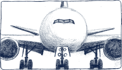
This three-part system can be compared to an airplane:
An airplane has a complex mechanical structure with many technical parts. But in order to fly, it also needs fuel—and for a successful flight, it must also have a pilot and air traffic control. All three are essential. Without any one of them, the plane can’t function properly.
When we return to thinking about higher brain function, we can see the same three essential parts:
- Mechanical Intelligence (Information Processing System)
This includes the ability to see, hear, understand what we see and hear, speak, remember, and more. In the airplane comparison, this is like the airplane’s machinery and technical systems—a very complex mechanical device.
But just as with an airplane, the machine alone isn’t enough to make it fly.
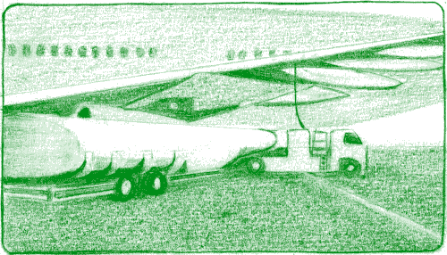
- The system that maintains alertness can be compared to the airplane’s fuel system.
Just like an airplane needs fuel in order to fly, the brain needs a certain level of alertness and energy to function. Without it, nothing can move forward—the brain simply can’t “take off.”
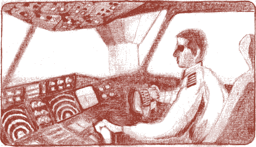
The executive function system—the part of the brain that plans, controls, and directs actions—is like the pilot and air traffic control.
If these don’t work properly, the “airplane” (your brain) won’t fly (work/function) at the right time, in the right direction—or might not fly at all.
Today, people often talk about cognitive functions when referring to higher brain functions in general. However, the term “cognitive” can be misleading. That’s because cognition specifically refers to knowledge and how we process information. Most people understand that higher brain functions involve much more than just information processing. So, while cognition is a part of higher brain function, it does not cover everything.
Another somewhat unclear term is “higher brain function” itself. In this context, “higher” doesn’t mean more important, more valuable, or even more complex. For example, controlling breathing is extremely important and not at all simple, but it isn’t considered a higher brain function in this context.
Because we need a shared term for complex brain abilities like thinking, language, initiative, decision-making, emotions, and emotional regulation and so on, we use the expression higher brain function. These abilities are especially characteristic of humans as a species.
Higher brain functions rely on widespread brain networks working together at the same time. The more these networks are required to work in sync, the more easily they can be disrupted. This is why the systems that control alertness and executive function—which use the broadest networks—are especially sensitive and vulnerable to damage.
In patients with brain tumors, disruptions or permanent damage to these networks can result not only from the tumor itself, but also from treatments such as surgery, radiation therapy, or chemotherapy. Seizures (epileptic episodes) can also damage the brain, and epilepsy medications may interfere with brain function. Corticosteroids (like dexamethasone) can impair brain function both directly and indirectly by affecting metabolism.
In the following sections we will go on to describe the symptoms caused by these types of damage or disruptions.
Impaired self-awareness of deficit – Lack of symptom awareness (anosognosia)
It can be difficult for a brain tumor patient to recognize and become aware of neurological and neuropsychological changes in his or her ability to function. The phenomenon is called impaired self-awareness of deficit.
The phenomenon is caused by damage and disturbances in the neural networks that maintain brain awareness (being aware or being conscious of oneself, self-awareness) and attention.
Patient’s close ones and caregivers may notice, for example, changes in reasoning and judgment, memory impairment, changes in behavior and emotional regulation, or mistakes in function that the patient is not aware of at all or only partially. A patient whose self-awareness is impaired may feel and think that his/her own ability to function is normal or almost normal. Giving feedback on symptoms and functional impairment is challenging in this situation. The patient’s ability to receive such feedback may be limited. In addition, this feedback might not lead to anything, either. The patient may understand or think he/she understands what is being said to, but nothing changes and nothing goes into practice. Often, only close ones see afterwards if the conversation was of any use. A patient with a lack of symptom awareness often has difficulty perceiving entities and contexts. He may not be aware of more complex situations and may not be able to report them.
Patient example: I met a younger brain tumor patient together with a specialized Neuro-oncologist. The patient’s tumor was mainly in the frontal lobes. It had been operated and radiated. The patient had had to move back to live with her mother because she could not manage living alone anymore. She had gained considerable weight and had stopped exercising almost totally.
At the office, we noted that the tumor was stable meaning that no treatment for the tumor was going to be started. The specialist talked at length to the patient and encouraged her to start exercising. The patient nodded understandingly, agreed completely and promised to try to start taking walks.
When the specialist left the room, I was there with the mother and the patient. I asked if the problem wasn’t that the patient did not get around to go for a walk. In addition, no amount of prompting and urging by the mother helped. The mother agreed fully. The patient sat passively there. We were then left to consider what kind of help the patient would need. The patient had just told the specialist that she was going to try taking walks. The next moment she agreed with her mother that she could not get it done. She was clearly not aware of her situation nor could perceive the relevant facts.
If she had been at the office alone and I had not been there either, the specialist would probably have been left with the impression that he had done his duty and the matter had been taken care of. In reality, nothing would have changed and the conversation would have been completely useless, possibly leaving the patient with a bit of anxiety without leading to anything else.
The extent and quality of symptom awareness problems are different among the patients because the location and extent of injuries are different in individual patients. If the patient is totally unaware of the symptoms and impairments the condition is called anosognosia2.( Greek: a=no, nosos= sickness, gnosis=knowledge). The lack of symptom awareness may also be partial, for example, so that the patient knows (intellectual awareness 1,3) that there are symptoms but is unable to anticipate (impaired anticipatory awareness 1,3) the actual functional limitations or problems resulting from the symptoms in order to function optimally. The patient may also have difficulty identifying how the symptom affects their own actions or reactions in the current situation or moment (emergent awareness3).
Symptoms of physical functioning are often better recognized and easier to be aware of than changes and deficiencies in cognitive or information processing, or in behavior and emotional regulation spheres. This applies both to patients and the patient’s relatives and caregivers. In some situations, it is indistinguishable whether the patient’s response is merely a lack of symptom awareness or, for example, an impulsive response in that situation or, for example, the patient is simply not remembering his or her functional limitations in the situation. It is noteworthy that a lack of symptom awareness due to brain damage is a different phenomenon than a partial or total denial of symptoms due to psychological reasons.
Healthcare providers may assume that patients are generally aware of their symptoms and functional changes, although this may not be the case. Deficient awareness of symptoms is a key problem for many patients and their close ones. This lack of awareness makes it difficult to assess one’s ability to function and goals in a realistic way. Lack of awareness also impairs both mental functioning and interaction with others (= psychosocial functioning). It would be important that the visits to the clinic should be made together with a close one who is well acquainted with the patient’s situation.
However, a large proportion of relatively healthy brain tumor patients visit the clinics alone. Functional problems that are evident in the patient’s own environment may then go completely unnoticed by the health care providers if the patient does not recognize them themselves and therefore cannot tell about them. It can also be that the healthcare personnel is not trained to assess patient’s higher brain functions, nor symptom awareness or it’s deficiencies.
Assessment of function and ability to work at the doctor’s office will also be incomplete and deficient if the patient is unable to describe in a realistic way his or her ability to function due to a lack of symptom awareness. If a close one or, for example, an employer describes problems with cognitive functioning that the patient does not recognize, it is always advisable to find out more about the patient’s symptoms and functional and work ability. Sufficiently extensive neuropsychological evaluation often provides relevant information. From the point of view of rehabilitation, the lack of symptom awareness is in itself a key target of rehabilitation measures1.
The following patient example illustrates impaired self-awareness of deficit:
The patient with a mostly frontal glioma had told the neurologist that he had no problems at work. In connection with a study, the neuropsychologist called the patient’s employer with the patient’s permission. The employer was relieved when someone finally asked about the patient’s ability to work. He told the neuropsychologist that the patient constantly forgot important things, mixed things up and made many mistakes with his work.
If somebody has to supervise, give directions and check the work all the time, the work output of the patient was naturally negative.
Self-awareness is assumed to be the result of the function of the neural networks and nodes in the frontal lobes of the brain, the lower parts of the parietal lobe, and the inner parts of the temporal lobes (Fig. 35 1 1)4. (Hearle L, Cocchi Brain Mapping 2015).
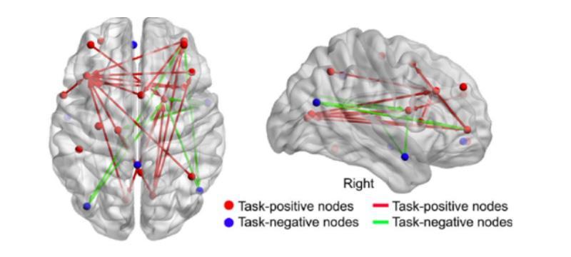
The main anatomical foundational structure is the fronto-parietal control network (FPCN) 4. This network consists of the frontal parts of the brain, particularly the right side –or rather these neural networks lie in the frontal, mostly right side parts of the brain (Fig 35 1 2). It is understandable that functional disturbances in this complex neural network are not due to a single, small lesion in the brain. In brain tumor patients there has not been much research about the FPCN network5.
Most oligodendrogliomas and other gliomas, i.e., supporting cell tumors, are located in the frontal, temporal, and parietal lobes. Consequently damage in FPCN and deficiency or lack of symptomatic awareness is a fairly common symptom in patients. Practical clinical experience from a follow-up clinic for brain tumor patients also supports this notion.
References:
1.Scherer M & Fleming J (2019) Awareness of deficits. In: JM Silver, TW McAllister & DB Arciniegas (Eds.), Textbook of Traumatic Brain Injury (3rd ed) (pp. 269-280). Washington: American Psychiatric Association Publishing. 2 Prigatano GP (2009) Anosognosia: clinical and ethical considerations. Curr Opin Neurol 22: 606–611. 3 Crosson B, Barco PP, Velozo CA et al (1989) Awareness and compensation in postacute head injury rehabilitation. J Head Trauma Rehab 4: 46-54. 4 Ham TE, Bonnelle V, Hellyer P et al (2014) The neural basis of impaired self-awareness after traumatic brain injury. Brain 137:586-597. 5 DeBaene W, Schouwenaars IT, Rutten GJT, Sitskoorn MM (2019) Task-evoked Reconfiguration of the Fronto-Parietal Network Is Associated With Cognitive Performance in Brain Tumor Patients. Brain Imaging Behav Aug 27.
Aivoväsymys –Brain fatigue (cognitive fatigue)
Normaalista poikkeava (patologinen) aivoperäinen väsymys
Aivojen vireystilan säätelyn ja ylläpidon toimintahäiriöihin liittyvä normaalista poikkeava kognitiivinen väsymys ja väsyvyys on huonosti tunnistettu ilmiö. Sitä voidaan kutsua aivoväsymykseksi, jota nimeä tässä käytetään. Seuraavassa käydään läpi sen piirteitä ja syntyä.
Normaalista poikkeava (patologinen) väsymys eli aivoväsymys on yleinen oire monissa neurologisissa sairauksissa, myös aivokasvaimissa1,2,3. Tällä aivoväsymyksellä tarkoitetaan sellaista ruumiillista (fyysistä) tai kognitiivista eli aivojen tiedonkäsittelytoimintoihin ja muuhun nk. korkeampaan aivotoimintaan liittyvää väsyvyyttä ja uupumusta, joka ei ole suhteessa fyysisten tai henkisten toimintojen tosialliseen kuormittavuuteen, on jatkuvaa ja haittaa toimintakykyä1,3. Aivokasvainpotilaiden väsymysoireita käsittelevässä melko tuoreessa katsausartikkelissa2 tällaista poikkeavaa väsymystä raportoi 39 – 77 % glioomapotilaista.
Aivokasvainpotilailla tätä aivoväsymystä ilmenee erityisesti kasvainhoitojen yhteydessä2. Väsymys helpottuu ja menee ohi osalla sairastuneista, mutta saattaa jäädä eriasteisena pitkäkestoiseksi. Aivoväsymyksen syitä2,3 voivat olla kasvaimen ja leikkausten mahdollisesti aiheuttamat vauriot ja häiriöt aivoissa, säde- ja solusalpaajahoitojen tai lääkitysten mahdolliset haitta- tai yhteisvaikutukset aivoissa ja elimistössä, psykologiset reaktiiviset tekijät, kivut ja unihäiriöt4,5,6. Fyysinen eli ruumiillinen väsymys on yleensä selkeästi ymmärrettävä tila. Aivojen vireystilan säätelyn7 ja ylläpidon toimintahäiriöihin liittyvä aivoväsymys (eli kognitiivinen väsyvyys6) on heikommin tunnistettu ilmiö, joka saatetaan tulkita virheellisesti esimerkiksi mielialasta tai motivaatiosta johtuvaksi tarmokkuuden ja jaksamisen puutteeksi.
Vireystilan säätelyn7 häiriöistä johtuvaa väsymystä voi esiintyä yhtäaikaisesti aivoperäisten unen häiriöiden kanssa, mutta sitä ilmenee myös potilailla, joilla ei ole unihäiriöitä tai unen puutetta6 eikä muuta3 väsyvyyttä selittävää tekijää. Aivoväsymykseen voi liittyä lisääntynyt unen tarve (pleiosomnia6). Herättyä vireystason nousu saattaa olla hitaahkoa ja päiväunien tai -levon tarvetta voi ilmetä.
Ihanteellinen vireystila on optimaalisen kognitiivisen toimintakyvyn eli kuhunkin tilanteeseen parhaan mahdollisen korkeamman aivotoiminnan perusta. Aivoväsymykselle on ominaista, että kognitiivinen kuormituksenkesto on rajoittunut, aivoväsymys aiheutuu normaalia vähemmästä ja herkemmin jopa yllättäen päivän aikana ja palautumisaika on tavanomaista pidempi6. Melko lyhytkestoinen ajattelutyö tai esimerkiksi kanssakäyminen ihmisten kanssa saattavat uuvuttaa. Aivoja kuormittavaa työtä on esimerkiksi kaikki lukeminen, myös SOME- kanavien, kirjoittaminen, elokuvien katselu ja luonnollisesti kaikki pohtiminen, ongelmanratkaisu ja vuorovaikutus jne. Vireystaso voi vaihdella päivän aikana normaalia enemmän tai olla riittävä vain osan aikaa päivästä. Päiväkohtainen vireystilan poikkeava vaihtelu on merkittävä toimintakyvyn haitta erityisesti työelämässä, vaikka osa päivistä olisi vireitä ja energisiä. Oireiltaan samantapaista väsymystä voi esiintyä myös syöpähoitojen yhteydessä tai niiden jälkeen henkilöillä, joilla ei ole mitään kasvainta aivoissa. Muut aivoja vaurioittavat tekijät, kuten vammat, tulehdukset ja verenkiertohäiriöt voivat luonnollisesti myös aiheuttaa samankaltaista aivoväsymystä.
Muuttuiko hänen persoonansa vai menettikö hän kyvyn käyttäytyä persoonansa mukaisesti?
Pohdintaa nk. persoonallisuuden muutoksesta – Reflections on so-called “personality changes”
Yhteenveto
Neuropsykologisten aivoperäisten henkisen toimintakyvyn häiriöiden, kuten käyttäytymisen ja tunnereagoinnin säätelyn häiriöiden, tulkitseminen persoonallisuuden muutoksiksi2 on kritiikille altis ajatusmalli. Pitäisikö pikemminkin puhua siitä, että potilas on menettänyt kykynsä käyttäytyä persoonansa mukaisesti? Kuvaisiko tämä ongelmia paremmin? Voiko olla kyse siitä, että potilas ei hermorataverkkojen häiriöiden tai vaurioden vuoksi aina kykene tulemaan ajatelleeksi-mitä missäkin tilanteessa tarvittaisiin?
Minkälainen ilmiö tämä niinkutsuttu persoonallisuuden muutos on?
Persoonallisuuden muutoksella5 aivojen vaurioitumisen tai aivotoimintojen häiriytymisen yhteydessä viitataan5 yleensä muutoksiin henkilön ulkoisessa käyttäytymisessä ja tunnereagoinnissa. Muutoksia voi myös olla yleisesti kyvyssä toimia, kommunikoida ja reagoida itselleen aiemmin tyypillisellä tavalla erilaisissa tilanteissa. Sairastunut saattaa esimerkiksi olla aiempaa puheliaampi tai toisinpäin niukemmin itseään ilmaiseva, aloitteettomampi ja laimeammin innostuva tai motivoituva, vaimeammin tilanteisiin reagoiva, vähemmän kiinnostunut läheisistä, joustamattomampi, kärsimättömämpi, herkemmin ärtyvä, estottomampi, impulsiivisempi, aiempaa taitamattomampi tulkitsemaan ja huomioimaan muiden tunteita ja reaktioita, herkemmin ahdistuva ja kuormittuva tai esimerkiksi tilannetaju ja kyky tehdä johtopäätöksiä saattavat olla puutteellisempia ja tunnesäätely vaihdella.
Tutkimuskirjallisuus ja potilaille sekä ammattilaisille tarkoitetut sivustot kuvaavat aivokasvainpotilailla esiintyvän näitä edelläkuvattuja persoonallisuuden1,2 muutoksiksi3,4,5 tulkittuja tiloja. Myös potilaiden läheiset ja potilaat itse kuvaavat samankaltaisia muutoksia. Muutokset voivat olla vähäisiä siten, että vain potilas tai potilaan läheisimmät havaitsevat ne. Toisaalta ne voivat toisessa ääripäässä olla niin voimakkaita, että ne kuormittavat potilasta ja läheisiä sekä haittaavat potilaan arkielämää, avustamista ja hoitoa. On myös tilanteita, joissa sairastunut ei itse kuvaa käytöksensä, toimintatapojensa, kommunikaationsa eikä reagointinsa muuttuneen, vaikka lääkärit tai läheiset toteavat niin tapahtuneen.
Persoonallisuuden muutoksiksi tulkittuja5 aivoperäisiä neuropsykologisia6 käyttäytymisen ja tunteiden säätelyn tai muita kognitiivisia häiriöitä ei ole kaikilla potilailla. Neuropsykologiset häiriöt voivat helpottua tai korjautua hoitojen, lääkityksen ja kuntoutuksen myötä. Huomattavaa on, että aivojen vaurioitumisesta johtuvat elimelliset tunnesäätelyn tai muut henkisen toimintakyvyn häiriöt ovat eri asia kuin sairastumisen aiheuttamat luonnolliset psyykkiset reaktiot.
Mitkä rakenteet aivoissa säätelevät käyttäytymistä ja toimintaa?
Aivojen etuosissa sijaitsevat otsalohkot6 hermoverkostoineen ovat keskeisiä käyttäytymisen ja tunteiden säätelyssä, sosiaalisissa taidoissa, harkintakyvyssä, impulssikontrollissa, tarkkaavuudessa ja keskittymisessä, kaikenlaisten toimintojen suunnittelussa, käynnistämisessä ja ohjauksessa sekä valmiudessa joustaa ja muuttaa omia toimintatapoja. Otsalohkotoiminnot ovat myös keskeisiä monimutkaisissa päättely- ja ongelmanratkaisutaidoissa ja kekseliäisyydessä, osassa keskeisiä muistitoimintoja sekä vaikuttavat motivoitumisessa. Vieläpä huumorin tuottaminen ja ymmärtäminen kuten myös ilmeiden ja äänensävyjen säätely ovat kytköksissä otsalohkojen toimintoihin.
Aiheellista onkin näin ollen pohtia, onko kyse kuitenkaan aivokasvainpotilaan persoonallisuuden muutoksesta2,5 vai itse asiassa elimellisistä kognitiivisten toimintojen ja käyttäytymisen tai tunteiden säätelyn neuropsykologisista6 häiriöistä, jotka näyttävät ulospäin ikään kuin persoonallisuuden muutoksilta, vaikka eivät sitä ole.
Potilasesimerkki perheyrityksestä: Mies oli ennen aivokasvaimeen sairastumista ollut keskeinen tätä palveluyritystä pyörittämässä. Kasvaimen ja sen hoitojen vuoksi hän joutui sairaslomalle. Viikolla hän selvisi hyvin, koska perhe oli joko yrityksessä työssä tai koulussa. Viikonloppu oli sitten kriisiä ja ongelmaa perheen kannalta. Mies lupasi osallistua johonkin yhdessä sovittuun ja ilmoittikin yhtäkkiä, ettei enää halua, ja poistui omaan huoneeseensa loukaten kaikkia. Kun perheelle selvisi, että potilaan joustavuus oli alentunut eli viikonlopuksi piti tehdä yhdessä sovittu aikataulu, tilanne helpotti vähän. Tämä tosin tuntui tylsältä perheestä, joka oli oppinut viettämään viikonloput ilman ennaltasovittua aikataulua. Lisäksi he oppivat, että potilaan kyky arvioida tilanteita ja omaa jaksamista oli puutteellinen ja että kaikkien perheenjäsenten läsnäolo oli kuormittavaa potilaalle. Ei sentähden, ettei hän isänä ja puolisona enää olisi pitänyt perheestään tai halusi välttää heidän seuraansa, vaan siksi, että hänen aivonsa eivät jaksaneet sitä viriketulvaa, jonka perheen kommunikointi ja puuhaaminen viikonloppuna aiheuttivat. Kun perhe ymmärsi tätä, he loukkaantuivat potilaaseen harvemmin ja osasivat säädellä potilaan kuormitusta paremmin. Lisäksi he oppivat, ettei tarvinnut pohtia potilaan persoonallisuuden muuttuneen, vaan voitiin miettiä eri tilanteita muuttuneen suorituskyvyn, kuten lisääntyneen kuormittuvuuden tai joustavuuden vähenemisen kautta.
Lähteet:
1 Kandler C, Zimmermann J & McAdams DP (2014) Core and surface characteristics for the description and theory of personality differences and development. Euro J Personal 28: 231–243.
2 Ojanen M (2019) Voiko persoonallisuus muuttua? Jyväskylä: PS-kustannus.
3 https://www.thebraintumourcharity.org/
4 Gempt J, Bette S, Albertshauser J, et al. (2018) Personality Traits in Patients with Neuroepithelial Tumors – A Prospective Study. Sci Rep 8:17055.
5 Jenkins LM, Drummond KJ & Andrewes DG (2016) Emotional and personality changes following brain tumour resection. J Clin Neurosci 29:128-132.
6 Goldberg, E (2001) The Excutive Brain: Frontal Lobes and The Civilized Mind. New York, Oxford University Press